
Labeled Muscle Diagram Chart Free Download
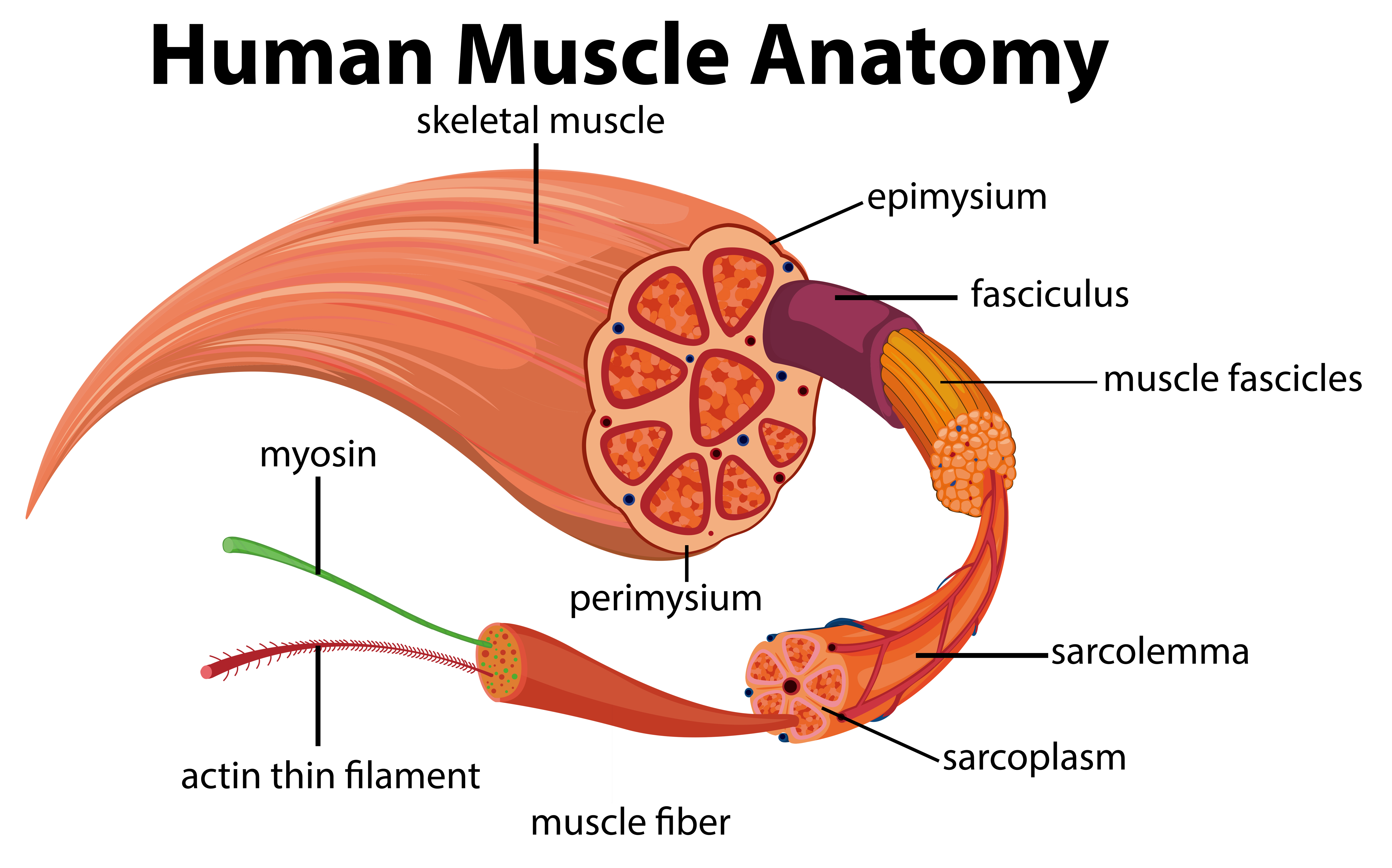
A typical myofiber is 2-3 centimeters ( 3/4-1 1/5 in) long and 0.05millimeters (1/500 inch) in diameter and is composed of narrower structures - myofibrils. These contain thick and thin myofilaments made up mainly of the proteins actin and myosin. Numerous capillaries keep the muscle supplied with the oxygen and glucose needed to fuel.
The Musculoskeletal System (Structure and Function) (Nursing) Part 4
Roll your mouse over any muscle in the diagram below to learn its name. You can click on any highlighted muscle to view a more detailed image of the muscle and a description of what it does.

human anatomy muscles labeled
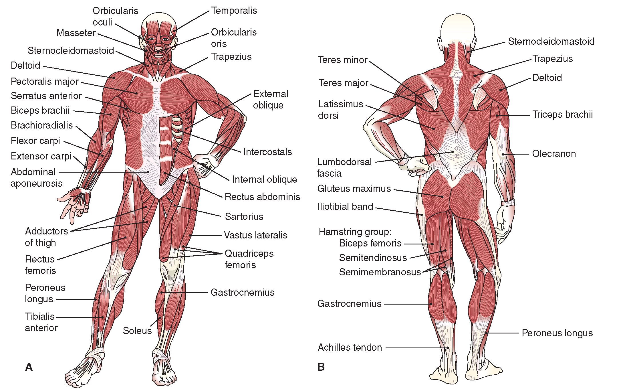
Chart of Major Muscles on the Front of the Body with Labels Major Muscles on the Front of the Body Last Updated On June 29, 2021 by Health Pages Team We have a lot of muscles in our bodies (literally, over 600). Muscles allow us to move and function. In general, they work in pairs.
labeled muscular system diagram Anatomy System Human Body Anatomy
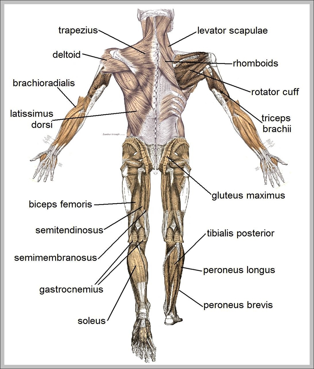
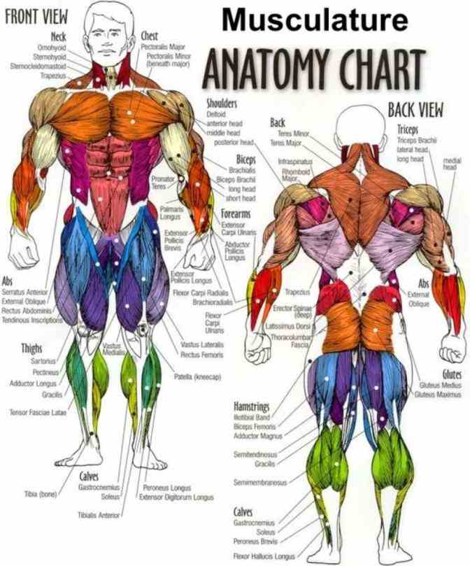
Muscle Charts of the Human Body For your reference value these charts show the major superficial and deep muscles of the human body. Superficial and deep anterior muscles of upper body Superficial and deep posterior muscles of upper body Anterior and posterior muscles of the upper arm Anterior and posterior muscles of the lower arm
Muscle Diagram Graph Diagram
Each skeletal muscle is an organ that consists of various integrated tissues. These tissues include the skeletal muscle fibers, blood vessels, nerve fibers, and connective tissue.
Images 05. Muscular System Basic Human Anatomy
Browse our selection of muscle models and charts, including the bestselling muscular system posters and muscle group chart. Home By Body Area Muscles Filters Brand 3B Scientific (37) Anatomical Chart Company (3) Anatomy Lab (2) AnatomyStuff (120) Denoyer-Geppert Science Company (1) Erler Zimmer (18) ESP Models (2) GPI Anatomicals (8)

Pin on Muscular System
Muscular system anatomy and physiology Sliding filament model of muscle contraction Muscle contraction Neuromuscular junction and motor unit Osmosis Muscles high-yield notes offers clear overviews with striking illustrations, tables, and diagrams. Make learning more manageable.
Labeled Body Muscle Diagram
Learn about the three types of muscle as you use our 3D models to explore the anatomical structure and physiology of human muscles. And don't worry, we'll explain the naming of skeletal muscles, too. By: Tim Taylor Last Updated: Oct 10, 2021 2D Interactive NEW 3D Rotate and Zoom Anatomy Explorer HEAD AND NECK CHEST AND UPPER BACK

Labeled Muscle Diagram Chart Free Download
Muscular System Anatomy, Diagram & Function | Healthline human body maps muscular system Muscular The primary job of muscles is to move the bones of the skeleton, but muscles also.
Muscles Diagrams Diagram of muscles and anatomy charts HubPages
Textus muscularis skeletalis Synonyms: Striated skeletal muscle, Textus muscularis striatus skeletalis Muscle is defined as a tissue primarily composed of specialized cells /fibers which are capable of contracting in order to effect movement.

the muscular system (lesson 0386) TQA explorer
Muscle is one of the four primary tissue types of the body, and the body contains three types of muscle tissue: skeletal muscle, cardiac muscle, and smooth muscle ( Figure 10.2 ). All three muscle tissues have some properties in common; they all exhibit a quality called excitability as their plasma membranes can change their electrical states.

Major Muscle Groups Body muscle anatomy, Muscle anatomy, Human body
Muscle diagrams are a great way to get an overview of all of the muscles within a body region. Studying these is an ideal first step before moving onto the more advanced practices of muscle labeling and quizzes. If you're looking for a speedy way to learn muscle anatomy, look no further than our anatomy crash courses .
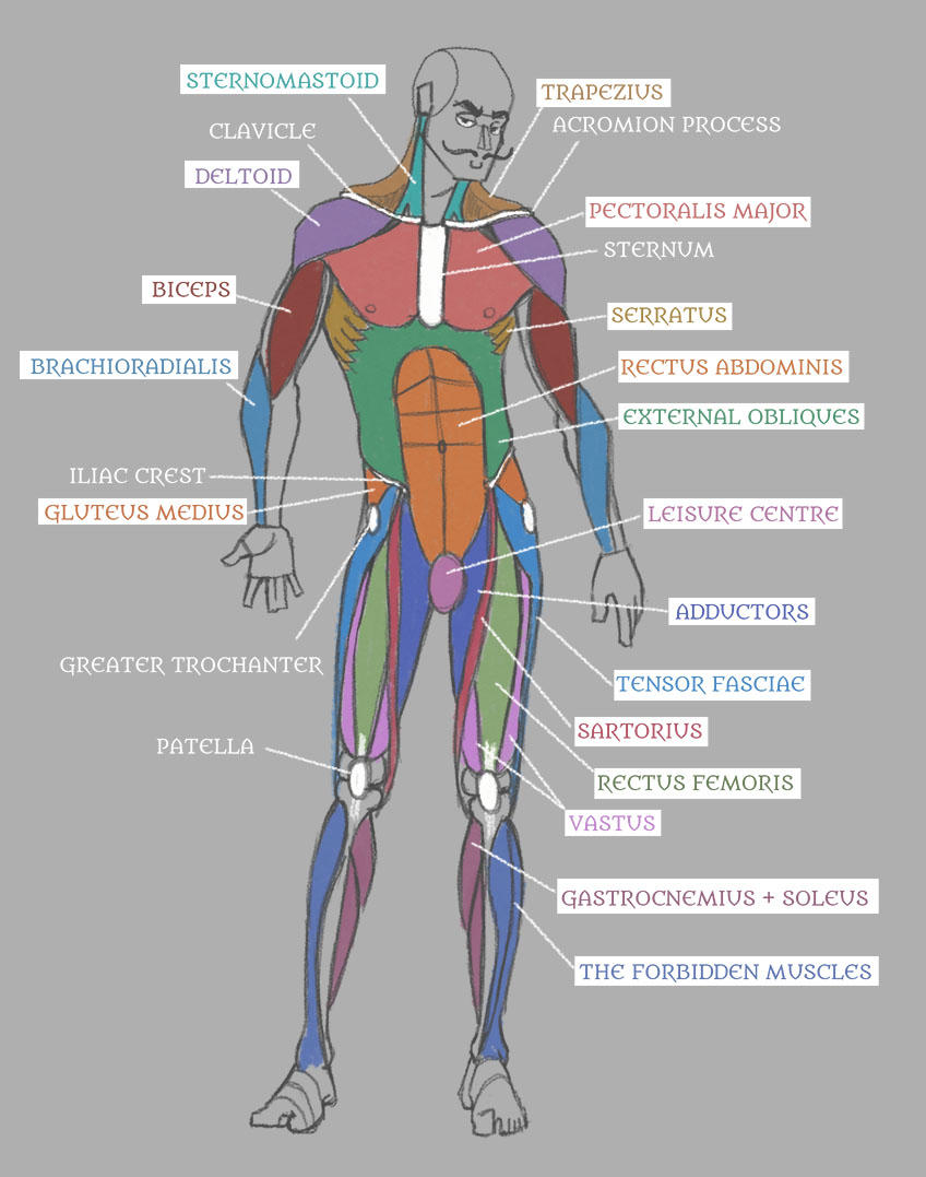
Human Anatomy Muscles with Labels! by Pseudolonewolf on DeviantArt
The musculoskeletal system comprises one of the body's major tissue/organ systems. The three main types of muscle tissue are skeletal, cardiac, and smooth muscle groups. [1] [2] [3] Skeletal muscle attaches to the bone by tendons, and together they produce all body movements.
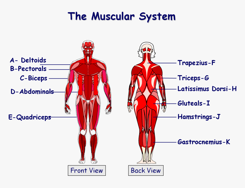
Diagram Of Muscles In The Body Amazon Com Human Body Muscle Anatomy
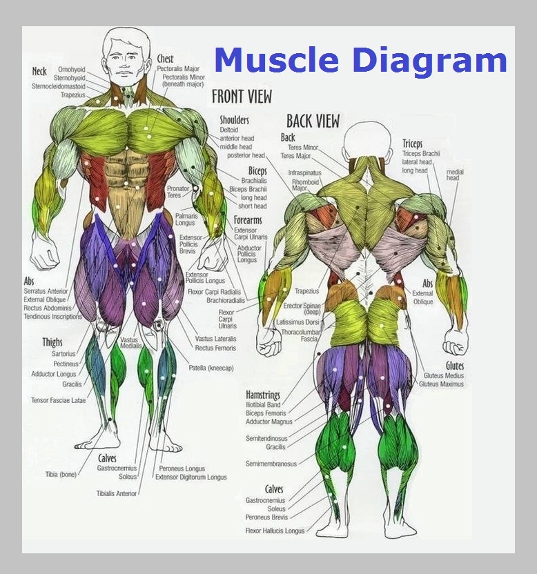
Last updated on October 27, 2019 Discover the muscle anatomy of every muscle group in the human body. Find the best weight lifting exercises that target each muscle or groups of muscles. You can click the links in the image, or the links below the image to find out more information on any muscle group.

The Muscular System Deep Layers, Back Laminated Anatomy Chart Human
The musculoskeletal system (locomotor system) is a human body system that provides our body with movement, stability, shape, and support. It is subdivided into two broad systems: Muscular system, which includes all types of muscles in the body. Skeletal muscles, in particular, are the ones that act on the body joints to produce movements.
Muscle Fiber Vector Art, Icons, and Graphics for Free Download
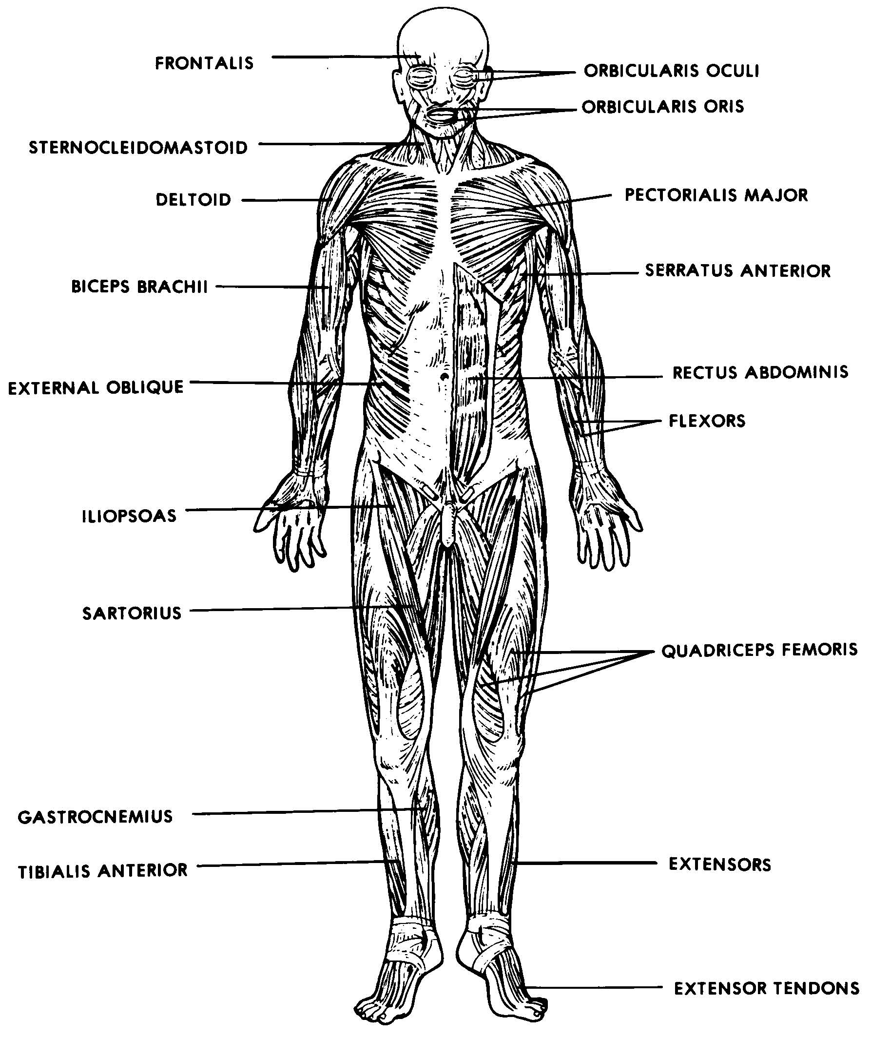
Human Anatomy - Front View of Muscles. Click on the labels below to find out more about your muscles. More human anatomy diagrams: back view of muscles, skeleton, organs, nervous system. Flex some.